


[Hepatobiliary [HB]] 14 Years/F, Incidentally detected hepatic mass
|
|
|---|---|
| Subspecialty | Hepatobiliary [HB] |
| Classification | Tumor |
| Difficulty | For resident |
| Modality | US, MR |
|
|
| 퀴즈 응모 기간 | 2025-10-10 ~ 2025-10-24 |
| Questions | What is your diagnosis? |
|
|
|
| ANSWER | |
|---|---|
| Answer | Focal nodular hyperplasia |
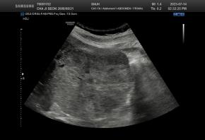
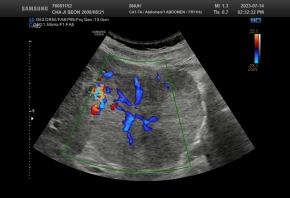
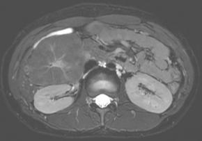
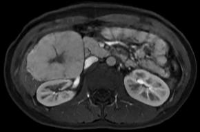
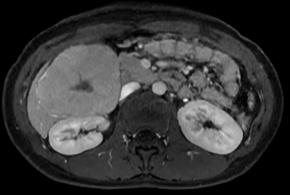
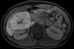
| Comments | This case report describes a hepatic tumor that was incidentally discovered in a pediatric patient with no underlying liver disease. On color Doppler imaging, there were findings suggestive of a central stellate scar, and the corresponding area demonstrated high signal intensity on T2-weighted MRI. Furthermore, on contrast-enhanced MRI, the lesion, excluding the central scar, showed homogeneous enhancement during the arterial phase and persistent enhancement in the portal venous phase with no evidence of washout. An additional finding, though not presented in the primary text, was homogeneous high signal intensity in the hepatobiliary phase (sparing the central scar). All of these findings are characteristic of focal nodular hyperplasia. |
|
|
| References | |
| Keywords | Focal nodular hyperplasia |
- 박영태 (2025-10-10 09:35:45)
- 모바일 홈페이지에서 정답이 보이는 문제가 있어요.
![]()
[04158]서울시 마포구 마포대로 53 마포트라팰리스 A동 304호
- 사업자등록번호 : 229-82-00887
- 대표자: 이재영
Copyright©by Korean Society of Ultrasound in Medicine
