



[Genitourinary [GU]] 26 Years/F, 복통 및 어지러움, 소화불량
|
|
|---|---|
| Subspecialty | Genitourinary [GU] |
| Classification | Tumor |
| Difficulty | For resident |
| Modality | US, MR |
|
|
| 퀴즈 응모 기간 | 2026-09-25 ~ 2026-10-09 |
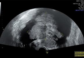
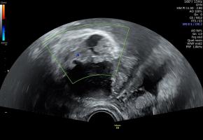
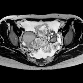
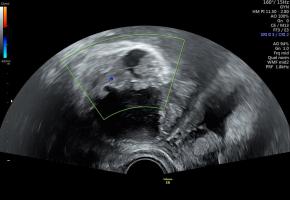
| Questions | 골반 초음파(B-mode, Color Doppler) 및 골반 조영증강 MRI(T2-weighted, Contrast-enhanced T1-weighted) 검사 영상이다. 외생성으로 자라나는 다발성 유두상 돌기(papillary projection)와 내부 혈류 신호가 관찰될 때, 가장 가능성이 높은 진단은? |
|
|
|
| ANSWER | |
|---|---|
| Answer | Serous borderline tumor |
| Comments | According to the updated IOTA consensus, a papillary projection is a solid component protruding into a cyst cavity with a height of at least 3 mm; papillary projections may arise from the cyst wall or a septum, and the largest projection should be measured using standardized dimensions. Previous studies have associated greater papillary height or number, blood flow, and irregular, confluent or disseminated morphology with borderline or invasive disease. Timmerman D, Valentin L, Testa AC, Froyman W, Landolfo C, Kotlarz A, et al. Terms, definitions and measurements to describe the sonographic features of adnexal tumors: updated consensus opinion from the International Ovarian Tumor Analysis (IOTA) Group. Ultrasound Obstet Gynecol 2026;67:702-724. Hassen K, Ghossain MA, Rousset P, Sciot C, Hugol D, Baddoura R, et al. Characterization of papillary projections in benign versus borderline and malignant ovarian masses on conventional and color Doppler ultrasound. AJR Am J Roentgenol 2011;196:1444-1449. |
| References | |
| Keywords | Ovary, Borderline tumor, Papillary projection, |

[04158]서울시 마포구 마포대로 53 마포트라팰리스 A동 304호
- 사업자등록번호 : 229-82-00887
- 대표자: 이재영
Copyright©by Korean Society of Ultrasound in Medicine
