



[Hepatobiliary [HB]] 71 Years/M, incidentally detected hepatic lesions
|
|
|---|---|
| Subspecialty | Hepatobiliary [HB] |
| Classification | Tumor |
| Difficulty | Rare case |
| Modality | US, CT, MR |
|
|
| 퀴즈 응모 기간 | 2026-10-25 ~ 2026-11-09 |
| Questions | What is the diagnosis? |
|
|
|
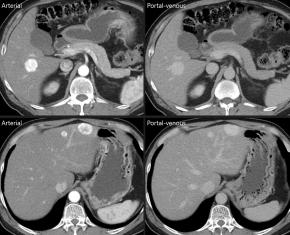
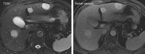
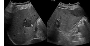
| ANSWER | |
|---|---|
| Answer | Meningioma metastasis |
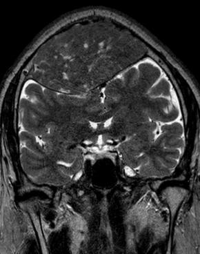
| Comments | The patient had a remote history of intracranial meningioma, and the hepatic masses were pathologically confirmed as metastatic meningioma. Extracranial metastasis from meningioma is extremely rare, with the lungs, bones, and liver being among the most commonly reported metastatic sites. Metastases may develop years or even decades after the initial diagnosis. Hepatic metastases may appear as well-defined, hypervascular masses on CT or MRI. However, imaging findings are nonspecific and may mimic hepatocellular carcinoma or other hypervascular metastases. |
|
|
|
| References | |
| Keywords | Rare hepatic tumor |

[04158]서울시 마포구 마포대로 53 마포트라팰리스 A동 304호
- 사업자등록번호 : 229-82-00887
- 대표자: 이재영
Copyright©by Korean Society of Ultrasound in Medicine
